 The Yoga Register

In classical kinesiology, the standing forward fold, known in postural yoga as Uttanasana, represents a closed-chain trunk flexion movement over a bilateral plantigrade base of support. While often categorized as a simple passive stretch for the posterior superficial line, the fold is an intricate balance between two primary articulation sites: the acetabulofemoral (hip) joints and the intervertebral joints of the spine. The mechanical success of the posture depends on the ratio of anterior pelvic tilt to spinal flexion, a relationship governed by muscle resting lengths, tendon compliance, and motor control.
When an individual descends into a forward fold, the center of mass moves anterior to the ankles, creating an external flexion moment that gravity amplifies. To maintain balance and prevent tissue overload, the nervous system must coordinate the rotation of the pelvis over the femoral heads while modulating the curvature of the lumbar, thoracic, and cervical segments. Analyzing this division of movement clarifies why standardized aesthetic cues often fail, and how uncalibrated movement patterns concentrate mechanical strain onto vulnerable passive connective tissues.
The Rhythm of Pelvic Tilt and Lumbar Flexion
Lumbopelvic rhythm describes the sequential and simultaneous contribution of lumbar spine flexion and pelvic rotation during forward bending. In an unconstrained, healthy descent, the movement does not occur in isolated compartments. Instead, early descent features an integrated combination of anterior pelvic tilt and progressive lumbar flexion. During the initial 45 to 60 degrees of forward movement, the pelvis rotates anteriorly at the acetabulofemoral joints while the lumbar lordosis gradually flattens. As the torso passes horizontal, pelvic rotation continues, while the lumbar spine moves into true flexion, reversing its resting inward curve into a slight outward kyphosis.
Research across occupational biomechanics indicates that a standard healthy forward bend requires roughly 55 to 65 degrees of hip flexion paired with 35 to 45 degrees of lumbar spine flexion. The precise ratio varies between individuals based on skeletal geometry, facet joint orientation, and passive soft-tissue tension. When this relationship functions properly, compressive and shear forces distribute across dozens of segmental joints, dissipating mechanical stress across the thoracolumbar fascia, intervertebral discs, and hip extensor muscle groups.
When pelvic tilt is restricted, the ratio distorts. If the pelvis rotates through only 20 or 30 degrees of anterior tilt because of mechanical or muscular constraints, the practitioner must generate the remaining displacement through the spine to bring the hands toward the floor. This forces the lumbar spine to reach its end-range flexion long before the torso approaches the thighs, transferring immense tensile loads to the posterior spinal ligaments and dramatic compressive loads to the anterior margins of the intervertebral discs.
| Movement Profile | Acetabulofemoral Flexion | Lumbar Flexion | Primary Tissue Restraint | Mechanical Risk Area |
|---|---|---|---|---|
| Balanced Rhythm | 55 to 65 degrees | 35 to 40 degrees | Shared muscular eccentric load | Evenly distributed force |
| Pelvic-Restricted Rhythm | 18 to 32 degrees | 48 to 58 degrees | Posterior spinal ligaments | L4-S1 intervertebral discs |
| Lumbar-Guarded Rhythm | 68 to 80 degrees | 10 to 20 degrees | Terminal hamstring tension | Proximal hamstring enthesis |
Hamstring Origin Mechanics at the Ischial Tuberosity
The hamstrings comprise three distinct muscles: the semimembranosus, the semitendinosus, and the biceps femoris long head. All three share a proximal origin on the ischial tuberosity of the os coxae. Because these muscles cross both the hip and knee joints, their length-tension relationship directly dictates pelvic mobility. In a straight-leg forward bend, the knee remains extended while the hip flexes. This position places the hamstrings under substantial passive tension, because both the proximal and distal attachment sites are pulled away from the muscle bellies simultaneously.
As the pelvis tilts anteriorly, the ischial tuberosities swing superiorly and posteriorly relative to the femoral shafts. This bony excursion directly increases the distance between the hamstring origin and the tibial insertions. If the hamstrings lack adequate resting length or exhibit elevated resting tone via protective neural guarding, they act as an immovable tether on the ischium. Once the hamstring reaches its maximal passive elastic limit, further anterior pelvic tilt halts. Any continuing descent of the torso must then originate entirely from segmental spinal flexion.
The mechanical stress at the ischial origin during this arrest is concentrated. When the hip is flexed to 90 degrees or beyond with an extended knee, the proximal hamstring tendon wraps around the lateral aspect of the ischial tuberosity. This anatomical wrapping creates a combination of high tensile stress along the longitudinal axis of the tendon and localized compressive stress where the deep tendon fibers press directly against the bone. In repetitive or prolonged end-range forward folds, this localized compressive-tensile shear can provoke insertional tendinopathy at the ischial enthesis.
Compressive Forces on Intervertebral Discs During Gravity-Assisted Bends
During standing postures, the gravitational force acting on the head, arms, and torso produces an external flexion moment around the lumbar spine. In an upright posture, the moment arm of the upper body relative to the L4-L5 and L5-S1 disc spaces is small, measuring only a few centimeters. As the trunk hinges forward into space, this moment arm expands substantially, reaching its peak horizontal distance when the torso is parallel to the floor. To prevent an uncontrolled fall forward, the posterior kinetic chain must generate an internal extensor moment of equal magnitude.
Classic in vivo intradiscal pressure measurements demonstrate this phenomenon clearly. Standing upright with a neutral spine generates an internal pressure of approximately 450 to 550 kilopascals in the L3-L4 nucleus pulposus. When an individual bends forward with straight legs and a rounded back, this pressure increases significantly, frequently surpassing 1200 to 1700 kilopascals. This surge occurs because the internal extensor force must be produced by the lumbar active vitality spinae, the multifidi, and the posterior passive ligamentous complex, all of which have relatively small lever arms and must pull with tremendous force to counteract the weight of the torso.
At terminal spinal flexion, an additional neurophysiological event occurs: the flexion-relaxation phenomenon. At approximately 80 to 90 percent of maximal lumbar flexion, the myoelectric activity of the lumbar active vitality spinae silences. The active muscular system shuts down, and the task of supporting the weight of the upper body shifts entirely to passive tissues: the supraspinous ligament, the interspinous ligament, the ligamentum flavum, and the posterior fibers of the annulus fibrosus. In this state, the lumbar intervertebral discs experience severe asymmetric loading, with heavy anterior compression wedging the disc space and driving the gelatinous nucleus pulposus posteriorly against the already tensioned posterior annular rings.
Knee Flexion: Angle Degrees and Hamstring Relief
To reduce disc compression and restore anterior pelvic tilt in individuals with hamstring tension, the distal constraint must be relieved. Introducing intentional knee flexion shortens the distance between the femoral condyles and the tibial and fibular insertions of the hamstrings. Because the semimembranosus, semitendinosus, and biceps femoris cross the posterior knee joint, bending the knee slacks these units across their distal end, releasing functional millimeters of excursion to be used at the proximal hip joint.
The degree of knee flexion directly alters pelvic orientation capacity:
- 0 to 5 degrees of knee flexion (Full Extension or Hyperextension): Hamstrings remain under maximal stretch. The ischial tuberosity is held taut. Anterior pelvic tilt is mechanically restricted unless the practitioner possesses exceptional baseline flexibility.
- 15 to 25 degrees of knee flexion (Micro-bend): Slacks the hamstring complex sufficiently to unload terminal neural and fascial tension. This allows an additional 12 to 18 degrees of anterior pelvic tilt in moderately stiff individuals, substantially delaying the onset of compensatory lumbar flexion.
- 30 to 45 degrees of knee flexion (Functional Hinge): Unloads the hamstring origin almost completely from passive end-range tension. The pelvis can freely rotate anteriorly to track the femur, aligning the trunk closely with the thighs and maintaining the lumbar spine in a near-neutral lordotic or flattened state, avoiding the flexion-relaxation phenomenon.
Flexing the knees also shifts the ground reaction force vector slightly posterior, moving the center of mass closer to the midfoot and heel. This shift decreases the required muscular effort from the calf complex and soleus, decreases sciatic nerve tension across the popliteal fossa, and permits the quadriceps to stabilize the base through active knee extension control without locking the tibiofemoral joint into terminal hyperextension.
Progressive Assessment Checklist for Hip Hinge Integrity
Evaluating whether a practitioner is hinging from the hips or merely flexing the spine requires an objective, progressive assessment. The following procedures establish the integrity of the pelvic-to-spinal movement ratio without relying on aesthetic markers like floor contact.
Palpating the Anterior and Posterior Iliac Spines
Position thumb pads on the Posterior Superior Iliac Spines (PSIS) and index fingers on the Anterior Superior Iliac Spines (ASIS). As the practitioner begins descending from an upright stance, monitor the spatial movement of these bony landmarks. In a structurally sound hip hinge, the ASIS and PSIS move simultaneously forward and downward in an arc around the femoral heads. If the PSIS remains static in space or drops downward while the torso descends, the movement is occurring entirely through lumbar flexion, indicating that the pelvic rotation has locked.
Applying the Three-Point Dowel Alignment
Place a wooden dowel along the practitioner's back, aligned along the sagittal midline. The dowel must maintain continuous contact with three anatomical points: the sacrum, the thoracic spine between the scapulae, and the back of the cranium. The practitioner flexes forward from the hips with soft knees. The assessment ends the moment the sacrum pulls away from the dowel (indicating lumbar hyperflexion) or the back of the head loses contact (indicating forward head compensation). Measure the angle of the dowel relative to the vertical axis at this cutoff point to establish their functional hip hinge threshold.
Executing the Wall-Block Distance Test
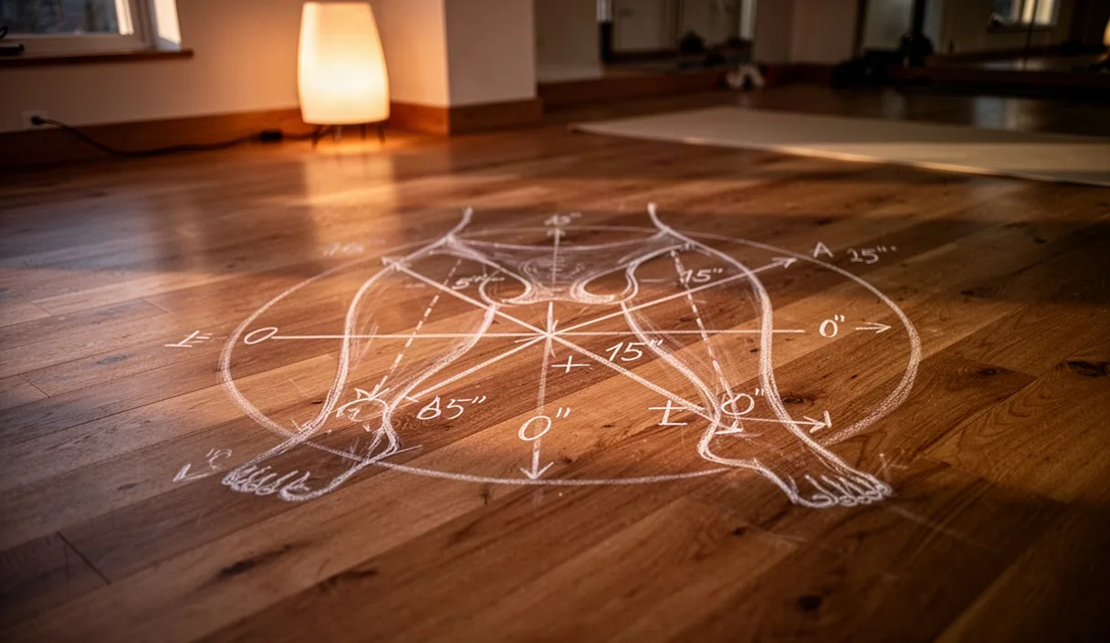
Stand with the heels placed exactly 15 centimeters away from a flat wall, feet parallel and hip-width apart. Position a light yoga block between the inner mid-thighs to maintain adductor engagement and prevent internal knee collapse. Keeping the spine in a neutral posture, cue the practitioner to fold forward by sending the ischial tuberosities back until the gluteal muscles make contact with the wall. If the knees shoot forward over the toes or the spine immediately rounds into kyphosis before the gluteal contact occurs, the practitioner lacks the posterior weight shift and anterior pelvic tilt mechanics needed for a safe standing fold.
Common mistakes
The most pervasive error in standing forward folds is prioritizing hand-to-floor contact over hip hinge mechanics. When reaching the floor becomes the primary objective, practitioners instinctively collapse the thoracic spine into maximum kyphosis, protract the scapulae, and drop the cervical spine into deep flexion. This generates the illusion of a deep fold while the hips remain stuck at 40 or 50 degrees of flexion, concentrating all kinetic energy directly into the interspinous ligaments and posterior disc margins of the lower back.
Another critical mistake is hyperextending the knee joints into genu recurvatum. Locking the knees backward wedges the anterior horn of the meniscus between the femoral condyle and tibial plateau while placing maximum strain on the posterior cruciate ligament and oblique popliteal ligament. Furthermore, hyperextension fixes the tibial insertions of the hamstrings at their furthest possible distance from the pelvis, solidifying the muscular brake on the ischial tuberosity and forcing the lumbar spine to compensate for the lost range of motion.
Practical Next Steps and Application
To apply these mechanics systematically in practice or teaching, implement a strict hierarchy of movement cues. Begin every standing forward fold with an active knee bend of at least 20 degrees. Instruct practitioners to maintain a long spinal axis, cuing axial extension through the crown of the head while consciously driving the sit bones upward and backward. Only after the anterior pelvic tilt reaches its individual limit should the remaining range of descent be accepted, allowing the spine to flex naturally and evenly across all 24 presacral vertebrae rather than hinging sharply at a single lumbar segment.
When working with practitioners who report active lower back discomfort, sharp posterior thigh pain, numbness, or tingling radiating below the knee, discontinue deep forward folding assessments immediately. These symptoms can indicate disc pathology, annular tears, or neural entrapment that require specialized diagnostic evaluation. Refer such individuals to an orthopedic physician or licensed physical therapist for a comprehensive clinical assessment before reintroducing gravity-assisted trunk flexion.