 The Yoga Register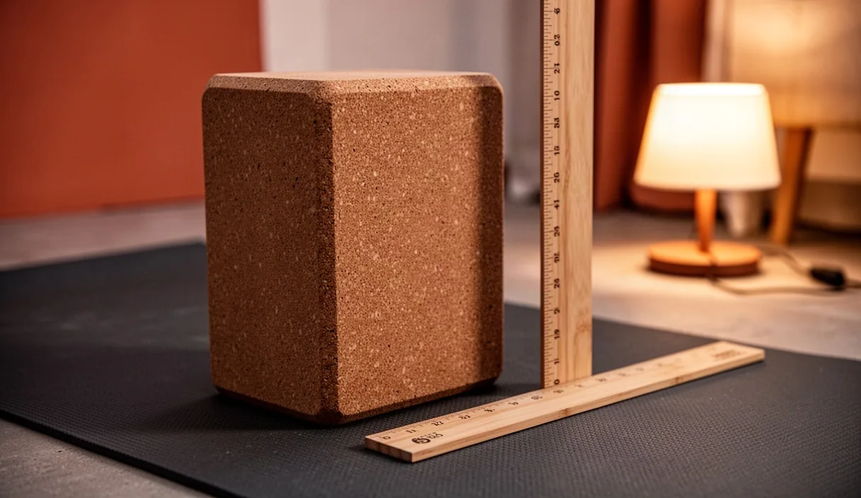

Chaturanga Dandasana, or Four-Limbed Staff Pose, is a low horizontal plank where the body is supported solely by the palmar surfaces of the hands and the plantar surfaces of the metatarsophalangeal joints. In this position, the human frame operates as a segmented cantilever beam. Because the center of mass sits roughly at the level of the lower abdomen, the gravitational vector exerts a downward force that generates substantial flexion torque at the lumbar spine, extension torque at the hips, and adduction and extension torques across the shoulder girdle. The upper extremity must generate sufficient counter-torque to maintain equilibrium without collapsing toward the floor.
The distribution of internal forces falls heavily on the shoulder complex, which lacks the bony congruency found in weight-bearing joints like the hip. Stability in this posture depends on balanced force couples across the glenohumeral and scapulothoracic joints rather than passive ligamentous end-stops. When the muscular architecture fails to stabilize these articulations, the mechanical load shifts to non-contractile tissues, including the glenoid labrum, the joint capsule, and the subacromial bursa. Understanding the mechanical demands of this posture allows practitioners and instructors to evaluate movement patterns objectively and apply load within functional tissue tolerances.
Anatomy of the Glenohumeral Joint at Ninety Degrees Flexion
The glenohumeral joint is a multiaxial ball-and-socket synovial joint characterized by a shallow glenoid cavity that accommodates less than one-third of the humeral head at any single point in its range of motion. In the bottom phase of Chaturanga Dandasana, the humerus rests at approximately 90 degrees of relative flexion or sagittal extension depending on the reference frame, with the elbow flexed to roughly 90 degrees and held close to the lateral torso. At this specific joint angle, the line of action of the ground reaction force runs superiorly and posteriorly through the forearm, driving the humeral head directly toward the anterior and superior margins of the glenoid fossa.
Passive stability at this threshold is minimal. The superior glenohumeral ligament and the coracohumeral ligament are slackened in this mid-range position, leaving the anterior band of the inferior glenohumeral ligament complex and the middle glenohumeral ligament to resist anterior translation. Consequently, dynamic muscular stabilizers must perform the primary restraint work. The rotator cuff musculature (supraspinatus, infraspinatus, teres minor, and subscapularis) must compress the convex humeral head into the concave glenoid fossa, a mechanism known as joint concavity-compression.
At 90 degrees of elbow flexion, the long head of the biceps brachii tendon experiences elevated tensile stress as it passes through the bicipital groove over the anterior humeral head. If the subscapularis fails to counteract the posterior pull of the infraspinatus and teres minor, or if the pectoralis major overpowers the posterior cuff, the humeral head translates anteriorly. This mechanical shear presses the biceps tendon and the anterior labrum against the static restraints of the joint, creating localized shear stress that exceeds normal resting thresholds.
Scapular Retraction versus Protraction in Horizontal Loading
The scapulothoracic joint determines the orientation of the glenoid fossa. In a prone horizontal loading state, gravity acts to drive the thoracic spine downward between the shoulder blades, promoting passive scapular retraction. However, unmanaged retraction shifts the glenoid fossa posteriorly and alters the subacromial space. The debate between conscious retraction and active protraction centers on how these positions alter the base of support for the humeral head and change the recruitment of the surrounding musculature.
Pure scapular retraction brings the medial borders of the scapulae within 5 centimeters of the vertebral column through the concentric contraction of the rhomboid major, rhomboid minor, and middle trapezius. While this stabilizes the shoulder blades against the rib cage in posterior pulling motions, it creates an unfavorable alignment during horizontal pushing. Unchecked retraction permits the anterior thorax to drop below the level of the humerus, placing the anterior shoulder capsule in an overstretched position under load. Conversely, maximal protraction, driven by the serratus anterior, draws the scapulae laterally around the thoracic wall, broadening the upper back. If driven into extreme kyphosis, maximal protraction closes the subacromial space, reducing clearance for the supraspinatus tendon.
| Scapular Position | Primary Active Muscles | Subacromial Clearance | Glenohumeral Joint Consequence |
|---|---|---|---|
| Passive Retraction | Rhomboids, Middle Trapezius (insufficient) | Moderately compromised | Anterior capsule experiences excessive tensile strain |
| Maximum Protraction | Serratus Anterior, Pectoralis Minor | Significantly reduced | Humeral head abuts the acromion process |
| Neutral with Lateral Wrap | Serratus Anterior, Lower Trapezius | Preserved (optimal) | Humeral head centered via dynamic concavity-compression |
The optimal mechanical objective is not maximal protraction, but rather a stable neutral position characterized by lateral scapular stabilization. In this state, the serratus anterior fires eccentrically or isometrically to prevent the medial border of the scapula from lifting off the posterior thoracic cage, an error termed scapular winging. Concurrently, the lower trapezius exerts a downward and inward vector on the scapular spine, preventing the upward migration of the shoulder blade toward the cervical spine and maintaining an open subacromial corridor.
Common Fault: The Anterior Shoulder Dip
The most structurally hazardous compensation pattern observed in Chaturanga Dandasana is the anterior shoulder dip. This fault occurs when the anterior aspect of the acromion process tilts downward and forward, causing the proximal head of the humerus to jut anteriorly relative to the anterior deltoid. Kinematically, this represents an uncontrolled combination of scapular anterior tipping, internal rotation of the humerus, and excessive extension of the glenohumeral joint past the plane of the torso.
Several underlying biomechanical factors contribute to this breakdown:
- Pectoralis minor tightness or over-dominance: The pectoralis minor originates on ribs 3 through 5 and inserts on the coracoid process of the scapula. Hyperactivity or shortening of this muscle pulls the coracoid process anteroinferiorly, causing the inferior angle of the scapula to lift away from the posterior ribs.
- Latissimus dorsi and triceps fatigue: As the primary extensors and adductors tire, the practitioner loses the capacity to keep the humerus parallel to the rib cage. The elbows flare laterally beyond 45 degrees, changing the force vector from a compressive load to a destructive torque.
- Rotator cuff fatigue: When the infraspinatus and teres minor can no longer hold the head of the humerus back within the posterior compartment of the joint, the humerus slides forward against the relatively weak anterior capsule.
When the shoulder dips below the horizontal line of the elbow, the humerus moves past 90 degrees of relative extension. This introduces an anteriorly directed shear force against the anterior labrum. Over time, repeated loading in this compromised alignment can irritate the long head of the biceps tendon and contribute to subacromial impingement syndrome. Individuals experiencing persistent discomfort during or after these loading mechanics should consult an orthopedic specialist or licensed physical therapist for a targeted structural evaluation.
Wrist Angle Variables and Ground Reaction Force
The wrist acts as the base of support through which the upward ground reaction force is transmitted into the radius, ulna, and upper kinetic chain. In a standard Chaturanga Dandasana, the hands are placed directly underneath or slightly posterior to the elbows, requiring approximately 85 to 90 degrees of passive wrist extension. For many individuals, achieving 90 degrees of wrist extension under direct compressive load compresses the dorsal structures of the carpus against the dorsal rim of the radius, leading to dorsal carpal impingement.
Ground reaction force vectors vary significantly depending on where the practitioner places their center of mass along the sagittal plane. If the heels push backward, the angle of the forearm relative to the floor becomes acute, tilting past 90 degrees so the elbows end up posterior to the wrists. This relieves pressure on the wrists by reducing the required extension angle to roughly 70 or 75 degrees, but it increases the moment arm between the center of mass and the glenohumeral joint, demanding higher contractile force from the anterior deltoids and triceps.
Conversely, if the practitioner shifts too far forward over the tips of the toes, the elbow angle drops below 90 degrees, and the wrist extension angle may exceed 95 degrees. This dramatically increases both the compression within the carpal canal and the shear stress across the carpal ligaments. Hand setup adjustments can mitigate these joint forces without diminishing the muscular challenge of the posture:
- Radial index finger alignment: Placing the crease of the wrist parallel to the front edge of the mat, with the index fingers pointing straight forward, distributes weight evenly across the metacarpal heads, reducing localized loading over the pisiform and ulnar carpal bones.
- The carpal arch engagement: Active flexion of the distal interphalangeal and proximal interphalangeal joints, sometimes described as gripping the mat, activates the flexor digitorum superficialis and profundus, which offloads direct compressive stress from the carpal tunnel structures.
- Forearm wedge utilization: Elevating the heel of the palm on a 15-degree wedge reduces the required dorsiflexion angle at the radio-carpal joint while maintaining proper humeral alignment.
Measurable Modifications for Insufficient Serratus Strength
The serratus anterior provides roughly 60 percent of the force required to keep the scapula anchored against the rib cage during prone upper-body loading. When a student cannot prevent scapular winging or anterior dipping, full horizontal loading exceeds their current tissue tolerance. Modifications must offer an exact, progressive reduction in the percentage of total body mass supported by the upper extremities, allowing the stabilizing musculature to adapt systematically.
Block Support Beneath the Coracoid and Pelvis
Placing two foam blocks on their highest or medium setting (typically 15 to 22 centimeters in height) beneath the anterior shoulders serves as an absolute depth stop. The blocks contact the clavicle and acromion before the humerus extends past the torso, mechanically preventing the anterior shoulder dip. An additional block positioned under the pelvis eliminates the lumbar hyperextension that frequently accompanies shoulder fatigue. This configuration allows the practitioner to isolate scapular stabilization with roughly 35 percent less load than the full posture.
Incline Loading Against a Wall or Elevated Surface
Adjusting the angle of the body relative to gravity directly alters the load experienced at the hands. The mechanical demands can be precisely scaled by manipulating the angle of inclination:
- Set the hand height: Place the hands against a stable wall or sturdy platform at chest height, stepping the feet back until the body forms a 45-degree angle with the floor. This orientation reduces the upper extremity load to roughly 40 percent of total body weight.
- Lower to contact: Flex the elbows to 90 degrees over an eccentric count of 3 to 4 seconds, verifying that the elbows remain tracked within 30 to 45 degrees of the torso.
- Assess stability: Check that the medial borders of the scapulae remain flush with the thoracic wall throughout the hold. Maintain the bottom position for an isometric hold of 8 to 12 seconds.
- Progress the angle: As capacity increases, lower the platform height to a bench (roughly 30 degrees of inclination) to increase the load to approximately 55 percent of body weight before transitioning to horizontal floor variations.
Knee-Supported Plank Progression
Lowering the knees to the floor shortens the lower lever arm of the cantilever system. This modification supports approximately 53 percent of total body weight, compared to the roughly 68 percent supported in a full toe-supported plank. The key execution variable is preserving a straight line from the knees through the pelvis to the crown of the head. If the practitioner hinges at the hips, the center of mass moves posteriorly, underloading the serratus anterior and failing to build the strength needed for the full expression of the pose.
Common Mistakes
Small errors in joint alignment can dramatically amplify localized joint stress in Chaturanga Dandasana. The following kinematic faults frequently compromise shoulder mechanics:
- Flaring the elbows past 45 degrees: Allowing the elbows to point outward horizontally places the humerus in significant internal rotation under load, impinging the supraspinatus tendon against the anterior under-surface of the acromion process.
- Dropping the head below the thoracic spine: Allowing the cervical spine to drop into passive flexion shifts the visual focus straight down, which reflexively encourages the thoracic spine to collapse and the scapulae to tip forward into anterior tilt.
- Descending past ninety degrees of elbow flexion: Lowering the shoulders below the level of the elbows creates an acute angle that drives the humeral head into extreme extension, stretching the passive stabilizers of the anterior shoulder capsule under maximum gravitational load.
- Holding the breath to stabilize the trunk: Relying on a closed-glottis Valsalva maneuver to generate trunk stiffness increases intrathoracic pressure unnecessarily, often causing involuntary muscle guarding around the neck and superior trapezius that disrupts healthy scapular depression.
Closing and Practical Progression Steps
Progressing the shoulder mechanics of Chaturanga Dandasana requires consistent neuromuscular patterning rather than hurried repetitions. To build balanced strength and protect the glenohumeral structures, incorporate the following diagnostic steps into practice sessions:
Baseline Assessment: Begin in a tabletop position on hands and knees. Slowly perform five scapular push-ups, moving solely between retraction and protraction without bending the elbows. Note whether the shoulder blades glide smoothly across the ribs or wing off the rib cage. If winging occurs in this quadruped position, horizontal loading in Chaturanga Dandasana will inevitably overload the static shoulder structures.
Volume and Load Management: Instead of performing multiple uncontrolled repetitions during flowing sequences, limit dynamic repetitions to three to five high-quality executions per session. Hold the isometric base of the modified posture (such as the knee-supported or incline variation) for 6 to 10 seconds per repetition. Focus on keeping the collarbones broad, the elbows hugged close to the rib cage, and the crown of the head reaching forward to elongate the axial skeleton.
Persistent Symptoms: Pain felt in the front of the shoulder, numbness or tingling extending into the fingers, or sharp pain on the dorsal aspect of the wrist are direct signs of joint overload. These symptoms require an immediate step back to modified variations or a temporary cessation of prone upper-body weight-bearing. When pain persists outside of practice, consult a certified physical therapist or medical professional for a comprehensive joint and soft-tissue evaluation.